The Facts Not Misconceptions About Ruptured Achilles Tendons
Overview
 A rupture of the Achilles tendon means that there has been either a complete, or partial, tear of the tendon which connects the calf muscles to the heel bone. Usually this occurs just above insertion on the heel bone, although it can happen anywhere along the course of the tendon. Achilles tendon rupture occurs in people that engage in strenuous activity, who are usually sedentary and have weakened tendons, or in people who have had previous chronic injury to their Achilles tendons. Previous injury to the tendon can be caused by overuse, improper stretching habits, worn-out or improperly fitting shoes, or poor biomechanics (flat-feet). The risk of tendon rupture is also increased with the use of quinolone antibiotics (e.g. ciprofloxacin, Levaquin).
Causes
A rupture occurs when a patient overstretches the Achilles tendon, an act which causes it to tear partially or completely. Achilles tendon ruptures can occur during athletic play or any time the tendon is stretched in an unexpected way.
Symptoms
If your Achilles tendon is ruptured you will experience severe pain in the back of your leg, swelling, stiffness, and difficulty to stand on tiptoe and push the leg when walking. A popping or snapping sound is heard when the injury occurs. You may also feel a gap or depression in the tendon, just above heel bone.
Diagnosis
Your doctor will ask you about your symptoms and examine you. He or she may also ask you about your medical history. Your doctor may ask you to do a series of movements or exercises to see how well you can move your lower leg. He or she may also examine your leg, heel and ankle and may squeeze your calf muscle to check the movement of your foot. You may need to have further tests to confirm if your tendon is torn, which may include the following. An ultrasound scan. This uses sound waves to produce an image of the inside of your leg. An MRI scan. This uses magnets and radio waves to produce images of the inside of your leg.
Non Surgical Treatment
The best treatment for a ruptured Achilles tendon often depends on your age, activity level and the severity of your injury. In general, younger and more active people often choose surgery to repair a completely ruptured Achilles tendon while older people are more likely to opt for nonsurgical treatment. Recent studies, however, have shown fairly equal effectiveness of both operative and nonoperative management. Nonsurgical treatment. This approach typically involves wearing a cast or walking boot with wedges to elevate your heel; this allows the ends of your torn tendon to heal. This method can be effective, and it avoids the risks, such as infection, associated with surgery. However, the likelihood of re-rupture may be higher with a nonsurgical approach, and recovery can take longer. If re-rupture occurs, surgical repair may be more difficult. 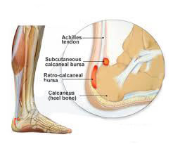
Surgical Treatment
The patient is positioned prone after administration of either general or regional anesthesia. A longitudinal incision is made on either the medial or lateral aspect of the tendon. If a lateral incision is chosen care must be taken to identify and protect the sural nerve. Length of the incision averages 3 to 10 cm. Once the paratenon is incised longitudinally, the tendon ends are easily identifies. These are then re-approximated with either a Bunnell or Kessler or Krackow type suture technique with nonabsorbable suture. Next, the epitenon is repaired with a cross stitch technique. The paratenon should be repaired if it will be useful to prevent adhesions. Finally, a meticulous skin closure will limit wound complications. An alternative method is to perform a percutaneous technique, with a small incision (ranging from 2-4 cm). A few salient points include: the incision should be extended as needed, no self-retaining retractors should be used, and meticulous paratenon and wound closure is essential. Postoperatively the patient is immobilized in an equinous splint (usually 10?-15?) for 2 weeks. Immobilization may be extended if there is any concern about wound healing. At the 2-week follow-up, full weight bearing is permitted using a solid removable boot. At 6 weeks, aggressive physical therapy is prescribed and the patient uses the boot only for outdoor activity. At 12 weeks postoperatively, no further orthosis is recommended.